Successful cochlear implantation produces a durable, reaction-free surgical site that allows the patient to use his/her implant system comfortably with a cosmetically acceptable appearance. The most frequently reported major and minor complications after cochlear implantation are those related to the incision and postauricular scalp flap. Ideal flap design that considers vascularity of the postauricular area, skull anatomy, device design, and proper surgical principles will prevent most complications from occurring. When complications do occur, treatment requires thoughtful and aggressive therapy to avoid further patient discomfort, morbidity, surgery, and dreaded device explantation. It cannot be stressed enough that prevention is the key.
Successful cochlear implantation produces a durable, reaction-free surgical site that allows the patient to use his/her implant system comfortably with a cosmetically acceptable appearance. Successful surgery also enables the patient to use an implant system to its fullest potential for accessing the sounds of speech and those of the environment. First steps in achieving an optimal surgical result require a plan of precisely locating the prospective implant site to facilitate interaction with the system’s processor and microphone. Adequate exposure of the surgical site requires that the surgeon develop a pedicled “flap” of skin and subcutaneous tissue. A well-planned flap provides exposure that facilitates safe mastoidectomy, and eliminates the risk of subsequent skin loss and device exposure. To ensure long-term coverage and device durability, the flap should be able to withstand the effects of pressure from the underlying device and compression from the overlying magnetically retained headset.
The most frequently reported major and minor complications after cochlear implantation are those related to the incision and postauricular scalp flap.1 Problems range in severity from minor wound dehiscences or infections to major loss of tissue necessitating removal of the device. In a study by Hoffman and Cohen,2 the overall rate of flap complications was 4.5%. Fifty-five percent of all flap complications were considered major and required follow-up surgery. Ideal flap design that considers vascularity of the postauricular area, skull anatomy, device design, and proper surgical principles will prevent most complications from occurring. When complications do occur, treatment requires thoughtful and aggressive therapy to avoid further patient discomfort, morbidity, surgery, and dreaded device explantation. It cannot be stressed enough that prevention is the key.
Prevention of scalp flap complications
Prevention of scalp flap complications after cochlear implantation begins with good incision and flap design. Ideally, the flap should have adequate blood supply and venous drainage, allow enough exposure of the operative site, and sufficiently cover the device. Blood supply to the postauricular area involves mainly 2 branches off the external carotid artery: the postauricular artery and the occipital artery (Figure 1). Venous drainage parallels the arterial supply.
Various incisions have been advocated for insertion of the cochlear implant . All have advantages and disadvantages. Classically, the anteriorly based, C-shaped flap has the advantage of providing complete coverage of the internal receiver-stimulator with borders that do not cross the implant. One disadvantage of this flap design is that it does not allow gravity based venous drainage, and flap edema may occur. Often associated with complications, anteriorly based flaps are contraindicated when there is a previous postauricular incision.
Inverted U-shaped and J-shaped flaps take advantage of the posterior arterial supply from the occipital artery. Because these flaps have the disadvantage of the incision crossing the electrode lead as it enters the mastoid cavity, it is necessary to create an anteriorly based musculofascial flap (ie, Palva flap) under the scalp to bolster electrode coverage. The patient may have postoperative numbness of an area of scalp superior to the horizontal arm.
Other incisions include straight, the “lazy S” incision, and the extended endaural incision. Gibson et al developed a 7-cm vertical entry route found to minimize scalp infection and device extrusion. The minimal access surgical route created by O’Donoghue and Nikolopoulos is a 3-cm oblique incision. Both offer the advantage of a minimal (or no) hair shave and a smaller incision. The 3-cm “keyhole” operation is limited to the use of a selected stimulator and may be more difficult to perform safely. Shorter incisions and less hair shave do seem to be beneficial in the pediatric population because it improves the esthetic quality of the procedure and reduces the psychologic trauma of the intervention. A recent retrospective study showed no evidence that minimal hair shave adversely affects the rates of wound complications. However, the value of any modification of wound preparation procedures should be considered carefully when a prosthetic device is to be placed.
Flap thickness must be incorporated in surgical planning. As Hoffman and Cohen warn, flaps that are too thick will impede transmission of electrical signals, while flaps that are too thin may erode under magnetic pressure. In younger children who have a thin scalp, elevation of the postauricular tissue in continuity with the skin flap may protect the flap from necrosis secondary to magnet pressure. Flap thinning is unnecessary in the pediatric population.
Although children were indeed once thought to be at higher risk for major and minor complications from cochlear implantation, past studies, in fact, suggested no significantly heightened risk in children. As for all otologic surgery in children, the surgeon should remember that the lack of development of the mastoid tip, narrow tympanic ring, and lack of subcutaneous tissue in infants and young toddlers place the trunk of the facial nerve just below the skin. An incorrectly placed incision or aggressive deep dissection of the inferior postauricular tissue may injure the facial nerve when it is unprotected by the mastoid tip.
Design of the postauricular skin flap should be tailored to the child’s head shape. In older children, the lateral skull is usually thick enough to permit creation of an adequate well for the receiver-stimulator. In younger children in whom the skull is much thinner, the bone is often drilled to the level of the dura, or a mobile island of thin bone can be created over the dura in the center of the well for protection. Alternatively, many surgeons make attempts to thin the skull to approximate, but not reach, the level of the dura. Such a conservative approach may reduce the likelihood of dural neovascularization and the risk of bacterial translocation as a precursor to meningitis. Retention sutures are often placed between the bone and dura.
Careful handling of the flap with proper moisture and hemostasis, and suture placement away from the surface of the implant are especially important. The wound should be closed in layers without undue tension, and a pressure dressing should be applied at the conclusion of the operation. Because of their high reactivity, chromic sutures should be avoided in closure. Intraoperative flap design and plans for device positioning are assisted by the use of a mock implant and mock behind-the-ear processor. The planned position for the receiver-stimulator may be marked through skin to bone using methylene blue in a medium bore needle. The flap is elevated to expose landmarks of the mastoid cortex (ie, the spine of Henle, linea temporalis, and the mastoid tip), and at least 3 cm of bone above and beyond the mastoid for locating the well to accommodate the device.
Treatment of minor scalp flap complications
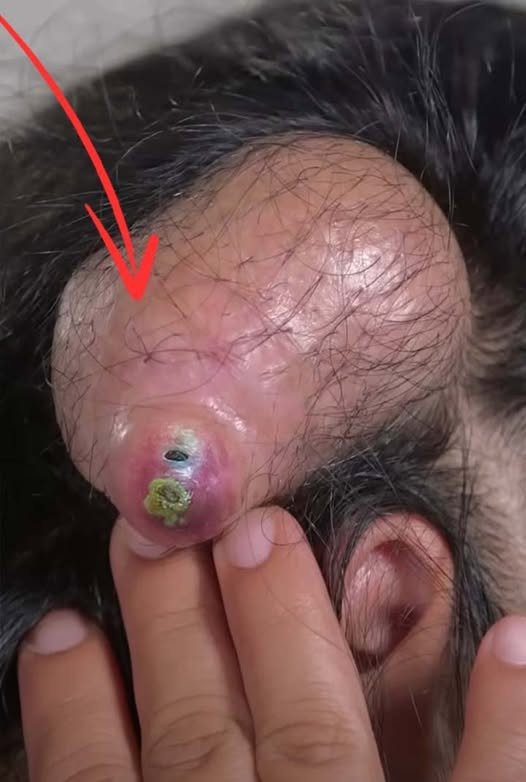
According to Cohen et al, minor scalp flap complications are those that require minimal treatment or no treatment to resolve. Luetje and Jackson labeled these the “nonsurgical” complications when they concerned the scalp flap. They are less frequently reported than major complications. Signs of flap infection should be immediately recognized and treated. Local symptoms and signs include erythema, warmth, and drainage and crusting at the incision site (Figure 4), which may be treated with topical and/or oral antibiotics. In adults, oral cephalosporins or fluoroquinolones are used. More persistent cases of infection should be treated with intravenous antibiotics, with consultation from infectious disease specialists to ensure that the blood prosthesis barrier is penetrated by the antibiotic. Aggressive therapy should be administered to prevent wound necrosis, which would then constitute a major complication requiring surgical intervention.
Figure 4. Early wound infection displaying erythema of incision site. (Color version of figure is available online.)
Wound dehiscence and/or delayed healing is rare but may occur more frequently in patients with underlying problems with healing (e.g., diabetics, smokers, immunosuppressed). When dehiscence occurs, meticulous wound care and aggressive antibiosis should be administered to prevent exposure of the device. Stitch abscess treatment involves local wound care, including possible incision and drainage with removal of the suture (Figure 5). Seroma/hematoma treatment depends on the size and rate of formation. Small collections that are not expanding may be treated conservatively with pressure and observation, and usually abate (Figure 6). Large or expanding lesions may need to be drained.
Treatment of major scalp flap complications
Flap necrosis is often the result of poorly planned and executed incisions or flap designs. Patients with previous postauricular or face-lift incisions should not be implanted using an anteriorly based, C-shaped flap because the blood supply to the flap may be inadequate. A “lazy S,” straight, or inverted U-flap or J-flap will allow survival of the flap. Infection and/or underlying inflammatory conditions (e.g., vasculitides) may also predispose to flap necrosis and problems with wound healing. There have been case reports praising the use of hyperbaric oxygen to speed recovery and healing, and even to “prepare” the bed for rotational flap.
Extrusion of the device (Figure 7) can result from local flap necrosis and infection transmitted from the mastoid. Paying particular attention to flap thickness intraoperatively and aggressively treating infection and minimizing comorbid conditions before surgery are effective preventative strategies. In cases in which adequately mobile, vascularized soft tissue in the postauricular area is unavailable, a rotation of the device or coverage with an extended flap, usually to a more superior location, may prevent device explantation. Other situations may require a rotational pericranial flap to fill the defect and enhance implant coverage.
Explantation is warranted when antibiosis and locoregional flap coverage fail to address a gathering wound infection. Reimplantation requires considerable attention to flap design. The skin over the anticipated implant site is often atrophic and must be handled carefully. The skin incision is through the original scar, and the flap is raised, with an attempt to maintain maximal thickness. The internal device well should be considered carefully. Often relocation of the device well is needed to provide coverage with adequate soft tissue.
Conclusion
The success of cochlear implantation has rested on its ability to enhance communication abilities of a large number of patients, while at the same time having minimal morbidity associated with the procedure. Major problems still occur, and scalp flap complications dominate these difficulties. Attention to detail, surgical planning and execution to prevent known mistakes, and aggressive treatment and recognition of postoperative complications may substantially reduce the risk of flap complication.